Michigan Technological University - India
In-Med Prognostics - Rajeshal Purushottam
- 2016
- Winner
- Venture
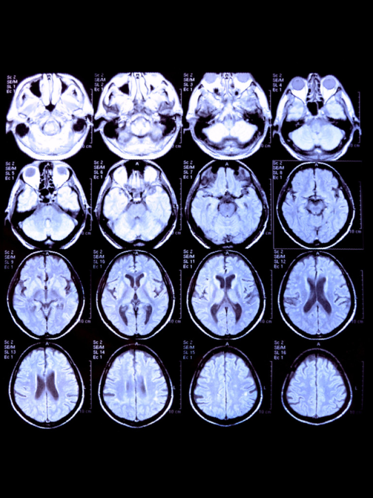
This is to provide objective quantitative neuroimaging reports at minimal price for early intervention in neurological illnesses. This product has the first-mover advantage in Indian and other emerging markets. Currently, there are few existing solutions to the challenges of neuroimaging analysis, if any. The difficulty lies in data generated with each image and the necessary objective inference by the physician.
Automated brain atrophy mapping software using volumetric MRI imaging for definitive quantitative analysis for neurodegenerative cerebral diseases. Early intervention is known to significantly enhance recovery in neurodegeneration patients of all ages and especially in young-onset dementia cases (Rosser.N et al, 2010). But there is a severe lack of cost-effective and reliable prognostic tools, especially in the emerging economies.
Existing prognostic solutions based on volumetric MRI imaging data are few and very expensive, catering only to developed economies. The only FDA cleared quantitative neuroimaging software is from a San Diego based company that charges about $89 for a single analysis. The cost makes this solution inaccessible for emerging markets such as India as well as other underserved populations in the developed economies.