Breaking the Wall of Deep Tissue Imaging
Breaking the Wall of Deep Tissue Imaging
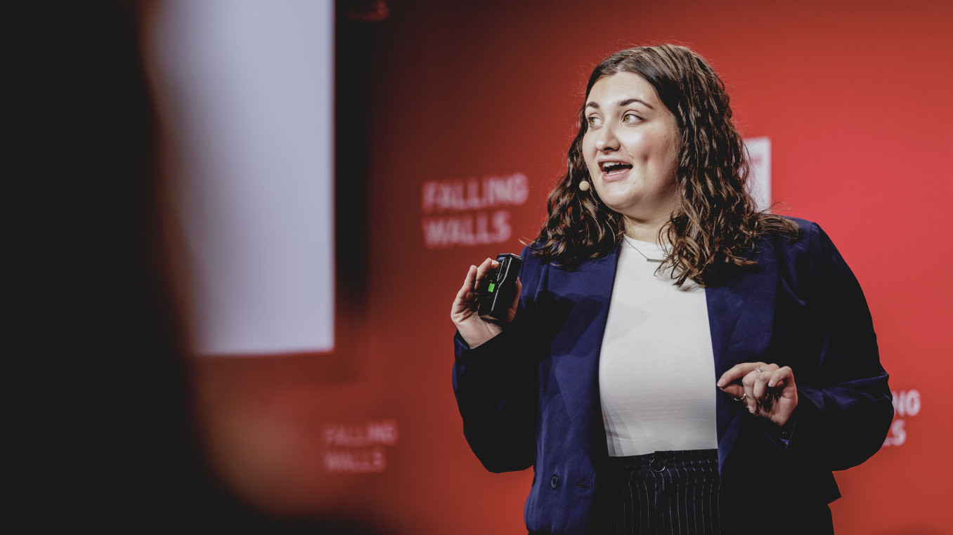
Global Call 2025 Finalist Interview: Life Sciences
David Maresca is an associate professor of Imaging Physics at Delft University of Technology in the Netherlands. He received his PhD in Biomedical Engineering from Erasmus University Medical Centre in Rotterdam, and his postdoctoral training from Institut Langevin in Paris and Caltech. David and his team pioneered imaging of genetically encoded sound-responsive proteins that function as the “GFP for ultrasound”. In 2025, his lab reported the development of sound-sheet microscopy, a method to visualise living opaque organs at the level of capillaries and cells.
Which wall does your research or project break?
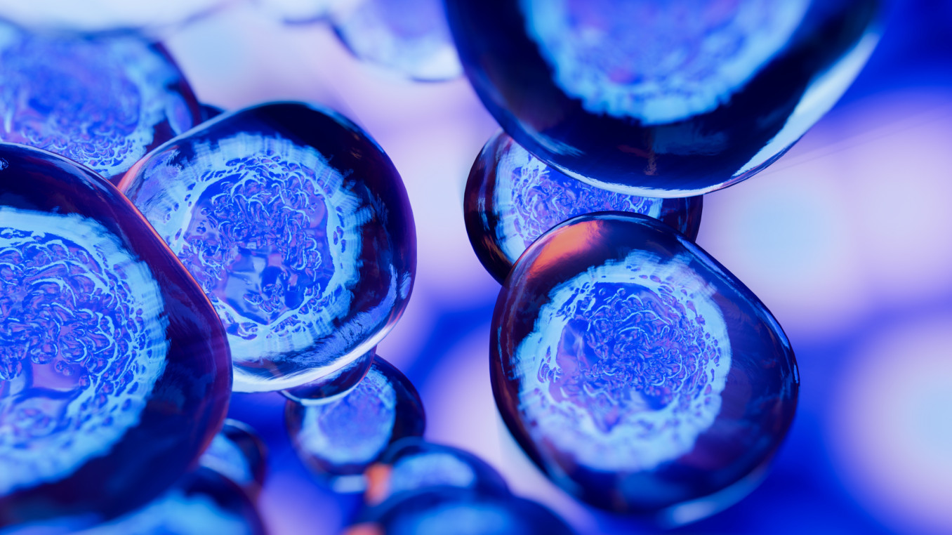
Visualising molecular and cellular processes occurring deep within living organisms is fundamental to our study of basic biology and disease. Currently, the most sophisticated tools available to dynamically monitor cellular events rely on light-responsive proteins which are difficult to use outside of optically transparent model systems, cultured cells or surgically accessed regions because of the strong scattering of light by biological tissue. In contrast, ultrasound enables the observation of living opaque organs in real-time and without any toxicity. However, its ability to detect cells was lacking. The recent introduction of sound-responsive proteins called gas vesicles allows ultrasound to connect directly to cellular functions such as gene expression.
To equip ultrasound with capabilities similar to those given to optical microscopy by fluorescent proteins, there is a need for fast high-resolution and volumetric ultrasound imaging methods capable of visualising acoustic reporter genes and acoustic biosensors. If this can be achieved, the resulting capabilities will allow researchers to explore cellular biology in vivo with unparalleled information content, resolution, coverage and translatability to biological research and clinical development.
In 2025, my laboratory introduced the concept of nonlinear sound-sheet microscopy, a method capable of detecting cells expressing sound-responsive proteins in thin tissue sections. Nonlinear sound-sheet microscopy can scan a cubic centimetre of biological tissue–the size of a sugar cube–with a resolution of 100 microns and a speed of 200 volumes per second. By introducing this method, we successfully broke the wall of deep tissue imaging of cells within living organisms.
What is the main goal of your research or project?
The discovery of the green fluorescence protein (GFP) and the parallel development of super-resolved fluorescence microscopy (2008 and 2014 Nobel Prizes) led to major breakthroughs in life sciences by enabling the visualisation of molecules on a cellular scale.
A major goal of my laboratory is to combine newly discovered biomolecules that scatter sound with dedicated ultrasound microscopy methods to enable the visualisation of cells on a tissue scale. If we are successful, the resulting capabilities will allow researchers to explore previously inaccessible cellular biology in vivo.
The physics of high-frequency ultrasound is ideally suited to in vivo cellular imaging by providing a combination of deep penetration (~1 cm) and high spatiotemporal resolution (~100 microns, 1 ms). Recent advances in imaging arrays have increased the field of view accessible from 2D (~1 cm x 1 cm) to 3D (~1 cm x 1 cm x 1 cm). Modern ultrasound hardware enables the acquisition of thousands of images per second but also real-time processing of this data. Finally, the discovery of genetically encoded gas vesicles, hollow protein nanostructures that scatter ultrasound waves, provides a platform for molecular engineering of acoustic reporter genes and acoustic biosensors. Together, these advances create the possibility for deep tissue imaging of biology at work.
A major upcoming frontier in imaging will consist of approaching single cell resolution with ultrasound (~10 microns) while maintaining a large field of view. To do so, super-resolution approaches grounded in wave physics will be investigated.
What advice would you give to young scientists or students interested in pursuing a career in research, or to your younger self starting in science?
My advice to a young scientist starting a career in research would be to develop “a prepared mind” or, in other words, to seek to obtain a deep understanding of the potential and intrinsic limitations of a given research field. At this stage, the specific research topic matters less than finding a welcoming mentor/suitable laboratory in which to learn and grow. In the long run, “a prepared mind” allows a seasoned scientist to "see" new opportunities when they present themselves.
Once the young scientist has more experience under their belt, my advice would be for them to engage in interdisciplinary research. Taking a sidestep is a chance to broaden your research horizon, to expand your network of peers beyond your first community and to discover the concepts, workflow, science vocabulary and limitations of an adjacent discipline.
My third piece of advice pertains to the consolidating phase of a research career. At this stage I would advise scientists to put some accepted norms and ideas to the test. If you have a hunch that an old theory is wrong but cannot prove it, you can test its pillars one by one. In imaging physics, the wavelength was thought to be the resolution limit. Medical textbooks still teach students that ultrasound waves cannot image through bone. These two examples were proven to be wrong.
Finally, I would say follow your curiosity and have fun!